
Figure 4.
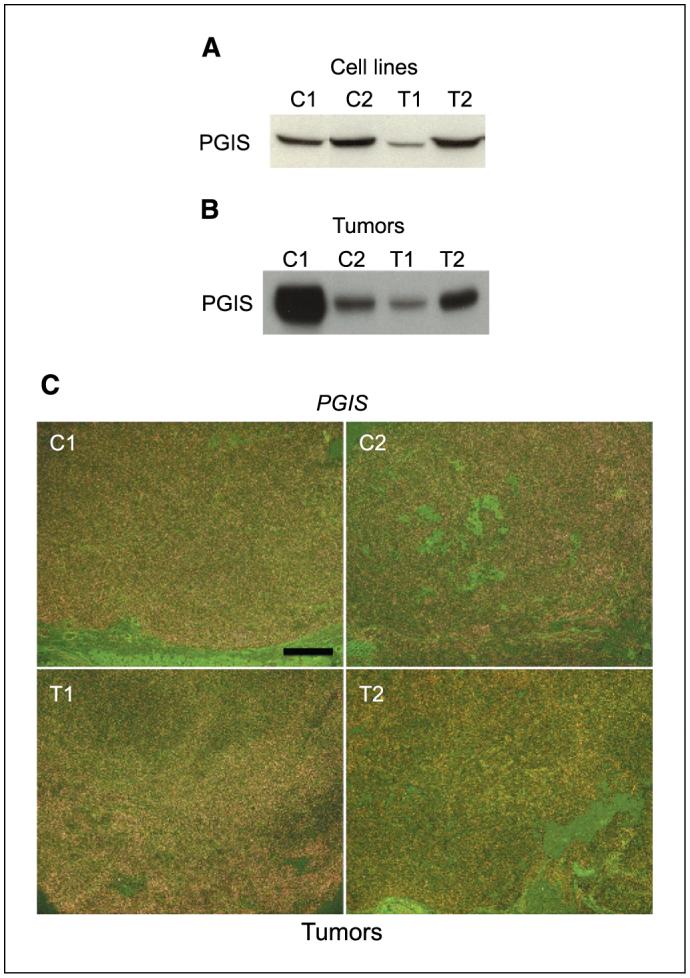
PGIS is expressed in OSE cells and tumors. A, Western blotting of PGIS in C1, C2, T1, and T2 OSE cell line samples. B, Western blotting of PGIS in tumor samples. C, in situ hybridization of Pgis in tumor sections. Representative darkfield photomicrographs of tumor sections show hybridization signals. Bar, 500 μm.