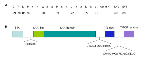
Figure 1.
Analysis of the primary structure of PSA proteins. A) Consensus sequence of the PSA LRR repeats. Numbers indicate the percentage of occurrence in the LRR repeats of the corresponding residues above. Residues in position #3, 7, 10, 13, 16, 18, 23(or 24) are always hydrophobic. B) Schematic representation of the PSA domain architecture. S.P: signal peptide; T/S: threonine/serine TM: transmembrane domain.

