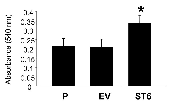
Figure 4.
Cell adhesion to collagen I. OV4 cells (P, EV, and ST6) were seeded onto culture dishes coated with collagen I, and binding was quantified using a standard crystal violet straining protocol. Data represent means and SEMs of three independent experiments run in triplicate. * denotes P < 0.05, evaluated by ANOVA.

