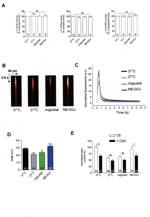
Figure 4.
Modification of local stimulation of caged Ca2+ in corrected F508del-CFTR CF15 cells. A Relative mRNA expression level of IP3R-1, IP3R-2, and IP3R-3 in different conditions compared to βActin mRNA expression. B Example of line-scan images acquired at 2 ms per line and 0.21 μm per pixel in CF15 cells treated (27°C, miglustat, NB-DGJ and uncorrected at 37°C in absence of extracellular Ca2+). C Average of the line-scan images in B expressed as normalized fluorescence in absence of extracellular Ca2+. D Histograms showing the amplitude of IP3Rs Ca2+ response in various experimental conditions as indicated. E Mean normalized area in each experimental treatment in absence or presence of 10 μM CsA. Sets of data were compared to the control CF15. Results are presented as mean ± SEM and the number of experiments is noted on each bar graph. ** P < 0.01, *** P < 0.001; ns, non significant difference.

