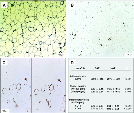
FIG. 1.
Histological characteristics of SAT and VAT. Slides of adipose tissue samples were stained with toluidine blue for the measurement of adipocyte area (A), anti–von Willebrand antibody for the evaluation of vessel density (magnification ×50) (B), and anti-CD45 (left) and anti-CD68 (right) antibodies for the quantification of inflammatory infiltrate (magnification ×100) (C). D: Results obtained in 35 patients (n) are expressed as means ± SD and were compared by paired t tests. All significant differences between SAT and VAT were also significant in unpaired t tests (P < 0.05). (Please see http://dx.doi.org/10.2337/db07-1812 for a high-quality digital representation of this figure.)

