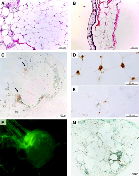
FIG. 4.
Connection between CAM vascular network and adipose tissue vessels. Viability of adipose tissue samples layered on the CAM was assessed by hematoxylin-eosin coloration of adipose tissue samples layered on CAM. Successfully grafted samples showing an angiogenic response had normal adipose tissue architecture (A), whereas adipose tissue degeneration was observed in samples without an angiogenic response (B). C: Staining with Hypoxyprobe at day 15 (incubated for 3 h at 21% O2 after an intravenous injection) showed a weak staining at the border between CAM and adipose tissue sample predominantly at the tip of the engulfment zone (see arrows) without staining inside the grafts. D: Adipose tissue samples layered on CAM from days 8 to 15 were stained with an antibody specific for chick erythrocytes. E: BrdU inside the samples was detected with an anti-BrdU antibody using samples that were collected 18 h after injection of BrdU in a CAM vein. An avian RCAS retrovirus-GFP was injected into the vitellin anterior vein of chick embryos on day 3. Adipose tissue samples were layered on the CAM on day 8. F: On day 15, fluorescence was observed in vessels formed around the samples, indicating that avian vessels had been the site of active angiogenesis. G: RCAS-GFP was detected in fixed adipose tissue samples stained with an anti-GFP antibody, indicating that cells of avian origin had infiltrated the vessels undergoing active angiogenesis inside the adipose tissue graft. (Please see http://dx.doi.org/10.2337/db07-1812 for a high-quality digital representation of this figure.)

