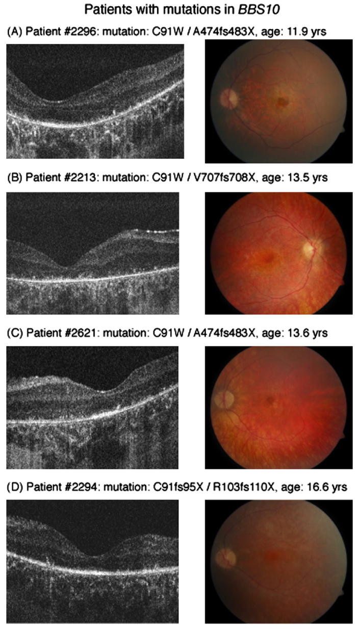
Fig. 3.

Horizontal Fd-OCT scans (5 mm) (left panel) and corresponding fundus photograph (right panel) of patients with mutations in BBS10. Maculopathy is early to moderate in patient #2296 (A) and #2621 (C) but absent in the other 2 patients (B) (D). ISL/OSL are identifiable in all but patient #2213 (B). Deposits above Bruch's membrane are visible in all patients (A–D, left panel).
