
Fig. 4.
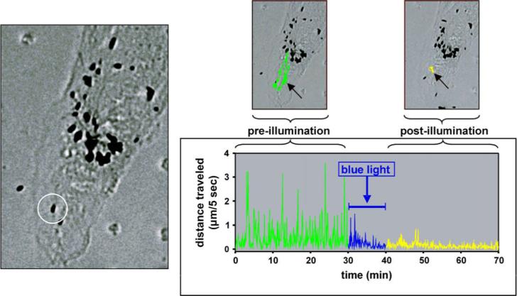
Effect of blue light treatment on the motility of phagocytized porcine melanosomes in ARPE-19 cells. Left: phase contrast micrograph showing a granule used for motility tracking (circled). Right: the granule's movement pre-illumination (green) and post-illumination (yellow) shown graphically and by colored traces superimposed on smaller micrographs. For the experiment, cultures were illuminated for 10 min with violet-blue light (400−410 nm at 4 mW/mm2) delivered via the epi-illumination system of the microscope using a protocol empirically determined to induce sub-lethal photic stress. To track granule movement, images were captured at 5 s intervals; image acquisition and data analysis were performed using Premier MetaMorph software. The blue light effect is quantified by comparing the total distance traveled over 30 min pre-illumination (188 μm) versus post-illumination (45 μm). For the granule illustrated here, motility decreased 76% on blue light treatment.