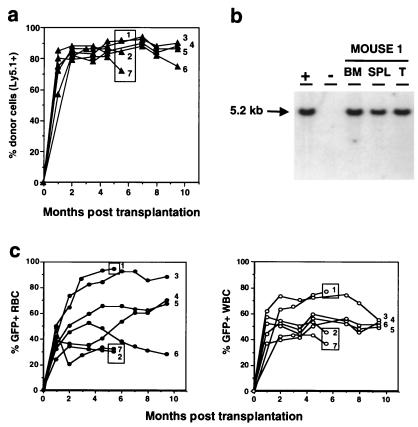
Figure 2.
Evolution of the reconstitution of peripheral blood cells with MSCVβ-globin/HS2/EGFP retrovirus-transduced, EGFP-positive cells. (a) Reconstitution kinetics of peripheral blood cells of recipient mice with donor-derived (Ly5.1+) cells. The proportion of Ly5.1+ peripheral blood leukocytes in each transplant recipient is shown. Boxed circles identify those animals that were killed at 5.5 mo posttransplantation for Southern blot analysis. (b) Southern blot analysis of a representative mouse killed 5.5 mo posttransplantation. DNA was isolated from bone marrow (BM), spleen (SPL), and thymus (T); +, DNA from the GP + E-86 MSCVβ-globin/HS2/EGFP viral producer shown to have X copies of integrated provirus; −, DNA from unmanipulated GP + E-86 packaging cells. DNA was digested with SacI and proviral integration assessed by probing with the EGFP gene. Left margin, Molecular size marker. (c) Reconstitution kinetics of the peripheral blood of recipient mice with EGFP-positive red (Left) and white (Right) blood cells.

