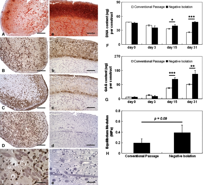
Fig. 3A–H.
SDSCs derived from conventional passage were contaminated with macrophages, inhibiting SDSC-based chondrogenesis. Histologic sections of 1-month constructs were stained: sulfated GAG (A/a; stain, Safranin O; magnification, ×100), collagen II (B/b; stain, immunostain; magnification, ×100), collagen I (C/c; stain, immunostain; magnification, ×100), and macrophage (stain, immunostain; magnification, D/d, ×100 and E/e, ×400). Sections A–E are for constructs engineered using SDSCs isolated by conventional passaging; sections a–e are for constructs engineered using SDSCs purified by negative isolation. Scale bars are 200 μm for A/a to D/d and 50 μm for E/e. (F) DNA content (μg/constructs), (G) GAG content (μg/constructs), and (H) equilibrium moduli of 1-month constructs are shown. Differences are indicated as follows: * = p < 0.05, ** = p < 0.01, and *** = p < 0.001. Data are shown as average ± SD (n = 4 constructs per group and time point).

