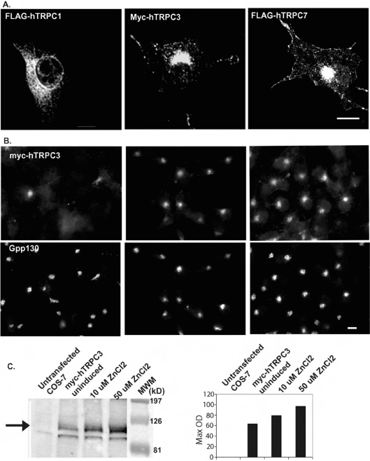
Figure 1. Expression of FLAG–hTRPC1, Myc–hTRPC3 and FLAG–hTRPC7 in COS-7 cells.
(A) COS-7 cells transiently expressing FLAG–hTRPC1, Myc–hTRPC3 or FLAG–hTRPC7 were processed for immunofluorescence microscopy using antibodies to the epitope tags. (B) A clonal cell line expressing Myc–hTRPC3 downstream of the metallothionein promoter (COS-hTRPC3-1) was grown for 48 h in the absence (uninduced) or presence of 10 or 50 μM ZnCl2 to induce increasing levels of expression. Cells were fixed and processed for immunofluorescence microscopy using a monoclonal antibody to the Myc epitope tag. (C) Untransfected COS-7 and COS-hTRPC3-1 cells were treated as in (B) and processed for immunoblotting. The immunoblot obtained using a monoclonal anti-Myc antibody is shown on the left. The filled arrow indicates the position of hTRPC3, which is absent in the untransfected cells. Increases in expression levels were quantified and are expressed as arbitrary units in the right-hand panel. Scale bars, 10 μm.

