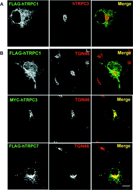
Figure 2. Co-expression of TRPC1 and TRPC3 does not alter their subcellular distribution.
(A) COS-7 cells were co-transfected with both FLAG–hTRPC1 and Myc–hTRPC3 constructs. Distribution of the tagged proteins was determined using a monoclonal anti-FLAG antibody and a rabbit polyclonal antiserum to TRPC3. Co-expression of the two constructs did not result in redistribution of either isoform. (B) hTRPC3 and hTRPC7, but not hTRPC1, co-localize with TGN46. Double-label immunofluorescence studies were performed on COS-7 cells transiently transfected with hTRPC1 (top panel) using a monoclonal antibody to the FLAG epitope tag and a sheep polyclonal antibody to a marker of the TGN, TGN46. Minimal overlap is seen as represented by a lack of yellow in the merged image. Cells transiently expressing hTRPC3 (middle panel) and hTRPC7 (bottom panel) were processed for immunofluorescence microscopy using a mouse anti-Myc epitope antibody, 9E10 (hTRPC3), or monoclonal anti-FLAG (hTRPC7) and sheep anti-TGN46 (middle panels). Overlap between hTRPC3 and both TGN46 and Gpp130 is shown in yellow in the merged images. Scale bar, 10 μm.

