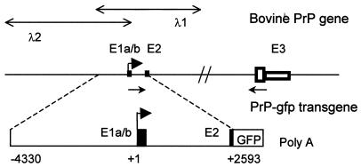
Figure 1.
Schematic representation of the bovine PrP gene and structure of the transgene used in this study. Exons (E) are numbered, and exon 1 and exon 2 are indicated by solid boxes. Within exon 3, the PrP ORF is shown by an open large box, and the mRNA 3′ UTR is represented by an open small box. The lengths of exons 1a, 1b, 2, and 3 are 53, 115, 98, and 4554 bp, respectively. The transcription initiation site is designated with a solid arrow (30). The positions of the two phages (λ1 and λ2) spanning the PrP gene are indicated. The primers used for the analysis of the 5′ UTR are shown by thin arrows (not to scale). The transgene encompasses 6.9 kb of the upstream sequences fused to the gfp reporter gene. GFP, gfp gene ORF; Poly(A), the simian virus 40 polyadenylylation signal. Positions are bp with respect to the transcription initiation site.

