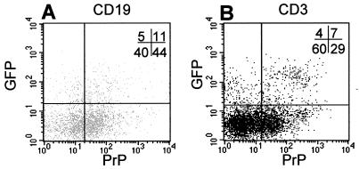
Figure 3.
Analysis of gfp and PrP expression from tg mice (L64) by fluorescence-activated cell sorter on peripheral blood leukocytes, gated for B (A) and T (B) lymphocytes. B cells were stained with R-phycoerythrin-conjugated anti-mouse CD19 antibody, T cells with Cy-chrome-conjugated anti-mouse CD3 molecular complex antibody (Becton Dickinson), and PrP-expressing cells with biotinylated anti-PrP antibody (8G8). The percentage of positive cells is indicated in the upper right corner.

