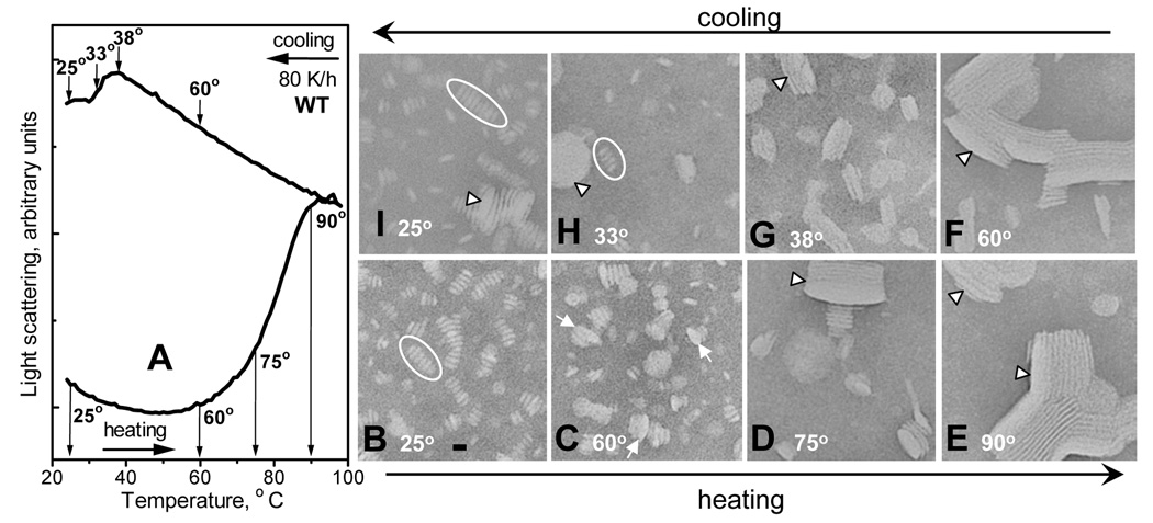
Figure 3.
Complexes of WT apoC-I:DMPC at various stages of thermal denaturation and reconstitution. Bar size is about 15 nm. (A) Light scattering data recorded of WT:DMPC disks during sample heating and cooling at 80 K/h from 20–98 °C; sample conditions are as in Fig. 1. (B–I) Electron micrographs of negatively stained WT:DMPC samples that were heated (lower row) and then cooled (upper row) to different final temperatures (indicated in the panels) that correspond to different stages of lipoprotein denaturation and reconstitution (indicated in panel A). At the final temperature, 1-naphthol was added to each sample (120 µM final concentration) to trap the reaction intermediates. Ovals in B, H indicate disk rouleaux, arrows in C indicate collapsed small unilamellar vesicles (that are thicker and larger in diameter than disks), and arrowheads in D–G indicate collapsed multilamellar vesicles.

