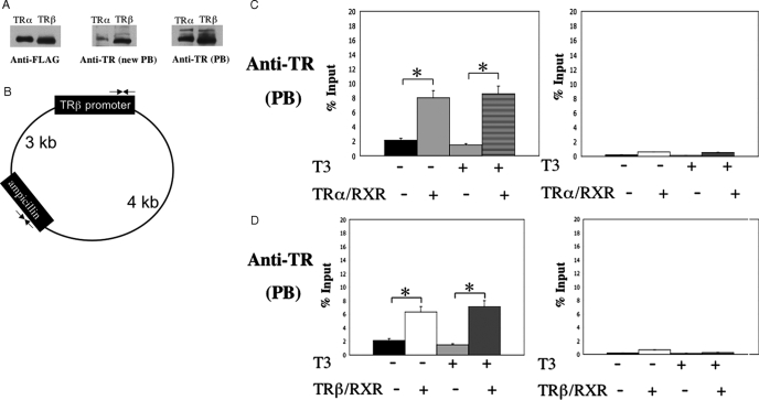
Figure 4.
Both TRs bind to the TRE in the chromatin in the frog oocytes. A, Western blot analysis shows that two independently generated anti-Xenopus laevis TRβ antibodies (PB and new PB) recognize both X. tropicalis TRα and TRβ, although the anti-TR (new PB) recognizes X. tropicalis TRβ a fewfold better than TRα. In vitro-translated FLAG-tagged X. tropicalis TRα and TRβ were subjected to Western blot analysis with the anti-FLAG, anti-TR (new PB), or anti-TR (PB) antibodies. B, Schematic diagram of the reporter plasmid containing the T3-dependent promoter of the Xenopus TRβΑ gene. Two sets (opposing arrows) of forward and reverse primers were designed for quantitative PCR analysis of the promoter region containing the TRE and a region in the ampicillin resistance gene, respectively. The primer sets are about 3 and 4 kb away from each other on the two sides of the plasmid, respectively. C and D, ChIP assays with anti-TR (PB) antibody show that chromatin-bound X. tropicalis TRα (C) and TRβ (D). Oocytes were injected with the reporter DNA and indicated mRNAs. After overnight incubation in the presence and absence of T3, the oocytes were isolated and subjected to ChIP assay with anti-TR (PB) antibody. The precipitated DNA was amplified for detection of the TRE region of the X. laevis TRβ promoter or the ampicillin resistance gene in the reporter vector. Note the enhanced signal of the promoter region but not the ampicillin resistance gene in the presence of TR/RXR both in the presence and absence of T3, showing the constitutive recruitment of TRs to the promoter.

