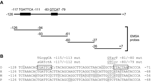
Figure 3.
A, Schematic representation of the proximal human FSHB promoter (−126 to +7) showing the sequences and relative position of two AP-1 cis-elements. The positions of the double-stranded DNA probes used in gel shift analyses are shown in the lower portion (see Table 1 for probe sequences). B, Alignment of FSHB/Fshb proximal promoter sequences, including putative AP-1 elements from sheep (O), human (H), mouse (M), and pig (P). AP-1 sites in the ovine promoter are boxed, and the putative AP-1 half-site in the murine and human promoters is underlined. Bases differing from the consensus are shaded. Mutations introduced into gel shift competitor probes and/or reporter constructs are shown at the top in lowercase/italics.

