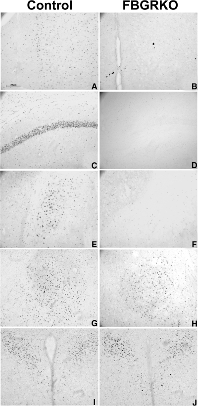
Figure 1.
Immunohistochemical verification. Immunostaining for GR in the medial prefrontal cortex (mPFC) (A and B), hippocampus (C and D), basolateral amygdala (BLA) (E and F), central amygdaloid nucleus (CeA) (G and H), and PVN (I and J) of control (A, C, E, G, and I) and FBGRKO (B, D, F, H, and J) mice. Note the absence of GR immunoreactivity in the mPFC, hippocampus, and BLA of FBGRKO mice. Importantly, the pattern and intensity of staining in the CeA and PVN is equivalent in FBGRKO mice and their littermate controls.

