
Figure 2.
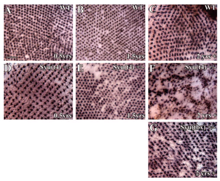
Age-dependent loss and maldistribution of blue cones in aging syu+/− zebrafish. Retinal whole-mounts from young (3–10 months pf; A, D), middle-aged (1.5 years pf; B, E), and old (2 years plus; C, F) wild-type (A–C) and syut4+/− (D–F) zebrafish were hybridized in situ with a blue opsin cRNA probe to identify the blue cones specifically. A precise blue cone mosaic was present in the wild-type retinas (A–C) and the youngest syut4+/− retinas (D). A few retinas from middle-aged syut4+/− fish were missing blue cones (E), whereas at least half of the oldest syut4+/− retinas were characterized by large regions lacking this cone photoreceptor type (F). Notice the blue cone-deficient regions are surrounding clusters of very densely spaced blue opsin-positive cells. (G) A disrupted blue cone mosaic, reminiscent of that observed in middle-aged syut4+/− fish, is evident in old syutbx+/− (see also Fig. 6). Scale bar, 25 μm.