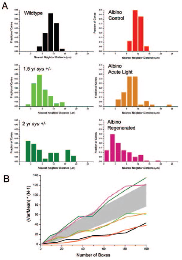
Figure 5.

Cone distributions in aged syut4+/− and light-damaged and regenerated alb zebrafish retinas are quantitatively similar. (A) NND histograms reveal a normal distribution of blue cones in wild-type (black) and control alb retinas (red) retinas. NND distributions are less “normal” in retinas from aging syut4+/− (light and dark green), light-damaged (orange), and the light-damaged regenerated retinas (pink). (B) Quadrat analysis of blue cone pattern in wild-type and control alb retinas (black and red lines, respectively) resulted in values of (var/mean)(N – 1) that are significantly lower (P = 0.05 criterion) than expected for a Poisson distribution, which is consistent with a highly regular pattern. Pattern analysis of the blue cones in middle-aged syut4+/− retinas (light green line) and alb retinas immediately after light-induced damage (orange line) showed values of (var/mean)(N – 1) that are not significantly different from those expected for a Poisson distribution (represented by shaded area), which are consistent with random patterns. Analysis of the blue cone pattern in an old syut4+/− retina and a light-damaged regenerated alb retina (dark green and pink lines, respectively) resulted in values of (var/mean)(N – 1) that are significantly higher than expected for a Poisson distribution, consistent with a clumped pattern.
