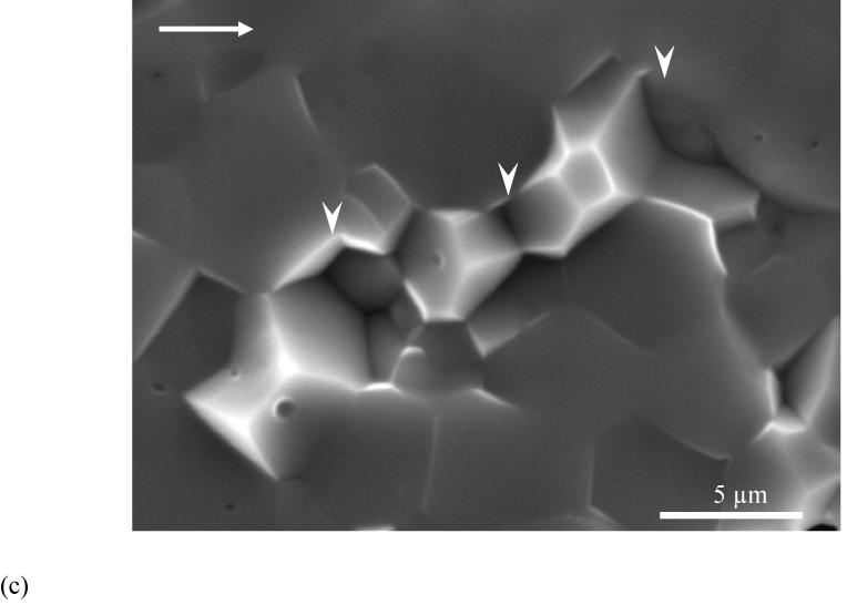
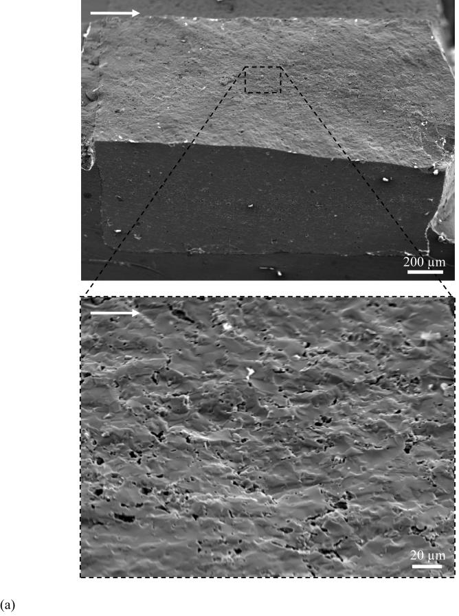
Figure 5.

Observations of crack extension in HAp.
(a) relatively smooth fracture plane of HAp compared to enamel in Figure 4
(b) a magnified view of fracture plane showing voids across the specimen thickness. The image was taken at the surface corresponding to steady state growth (ΔK∼ 0.2 MPa·m0.5).
(c) A typical high magnification image of fracture surface of the sintered HAp. Notice the surface is primarily uniform but does exhibit a few HAp crystals (white arrowheads) pullout.

