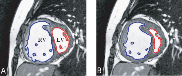
Figure 3.
Double-oblique steady-state free precession cine MR images (repetition time ms/echo time ms 3.3/1.65; flip angle 60 deg; section thickness, 6 mm; matrix, 256 × 150). End-diastolic (a) and end-systolic (b) cardiac short axis slice in PAH patient. The endocardial boundaries of the right ventricle (RV) and left ventricle (LV) are traced for calculation of end-diastolic volume (EDV) and end-systolic volume (ESV). EDV and ESV were calculated by summation of the product (area × slice distance) for all slices. SV is then given by SV = EDV-ESV for RV and LV.

