Figure 1.
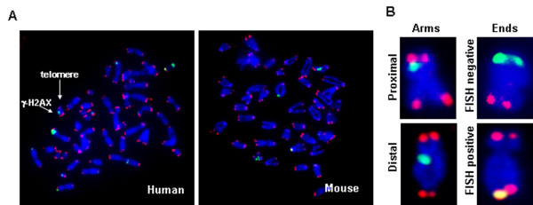
γ-H2AX immunostaining on metaphase chromosomes. (A) Typical metaphase spreads of human (left) and mouse (right) fibroblasts stained for γ-H2AX (green) and telomeric DNA (red). (B) Scoring of foci as along the chromatid arms, proximal or distal to the telomeres, or on the chromatid ends, fluorescence in situ hybridization negative or positive.

