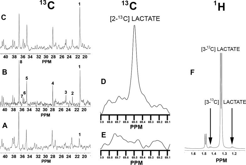
Figure 2.
Three representative 13C NMR spectra are shown for Perchloric Acid (PCA) Extracts from slices taken from the superfusion chamber at different times: (A) before hypoxia-ischemia; (B) at the end of hypoxia-ischemia; (C) after four hours of recovery following hypoxia-ischemia. Resonances for particular metabolites are numbered: 1) lactate C3, which is substantially increased in B and C relative to A ; 2) β−hydroxybutyrate C4; 3) GABA C3; 4) glutamate C3; 5) glutamate C4; 6) succinate C2, C3; 7) GABA C2; 8) taurine. (D) a different portion of the same spectrum shown in C, showing an increased resonance peak for C2 of lactate, which is barely discernable in (E). (F) A representative portion of a 1H spectrum at the end of hypoxia-ischemia. The large, narrow doublet at 1.33 ppm is from the methyl protons on the C3 of unlabeled lactate. When the C3 of lactate is labeled with 13C, two doublets occur as indicated by arrows instead of the narrow doublet at 1.33 ppm. In all spectra where 13C-glucose is was not used, the combined area of the two small doublets was 1.1% of the large central doublet, which is what occurs from natural abundance 13C.

