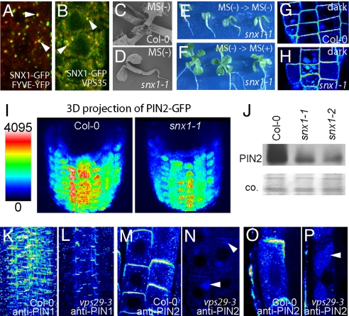
Fig. 3.
Regulation of PIN degradation by plant retromer components. (A and B) Merged images of colocalization (arrowheads) of SNX1-GFP (green) with the PI3P-binding domain FYVE-YFP (red) (A) and retromer component VPS35 (red) (B). (C and D) Two-week-old wild-type seedlings with normal growth on media without sucrose (C) and snx1 mutants arrested in growth (D). (E and F) Rescue of growth defects (E) by transfer on sucrose-containing media (F). (G and H) Substantial vacuolar targeting of the PIN2-GFP in the wild type (G) and even more pronounced in snx1 mutant (H) after dark treatments for 2 h. (I) Reduced PIN2-GFP levels at the plasma membrane in snx1 mutant. Three-dimensional animation of z-stacks (80 μM with 2-μM steps) was obtained. Roots were digitally tilted for outlook at the apical cell surface. False color code was used for PIN2-GFP intensity visualization. (J) Reduced total PIN2 protein levels in snx1 mutants by Western blot analysis. (K and L) Reduced PIN1 protein levels in vps29 (L) compared with wild-type seedlings (K) by z-stack analysis and maximum projection of the PIN1 immunolocalization (≈80 μM, 2-μM steps). (M–P) PIN2 immunolocalization in wild type (M and O) and vps29-3 mutants (N and P). PIN2 accumulation in vps29 was ectopically localized in vacuolar-like structures in meristematic (N) and in elongated root cells (P), suggesting enhanced degradation in vacuoles. Arrowheads depict PIN occurrence in vacuole-like structures. False color code depicts relative fluorescent intensity (I and K–P).

