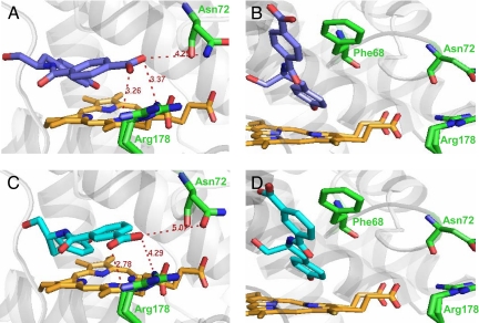
Fig. 5.
Modeled complexes of wild-type HRP with d-3 (A), l-3 (B), d-4 (C), and l-4 (D). For clarity, only the active site of the enzyme is shown with the heme moiety in orange, substrate in blue, and some mutated residues in green. Distances indicated are in angstroms. The Arg-178 residue is shown in a double rotamer configuration as it appears in the crystal structure (29); only 1 rotamer configuration was used in docking experiments. See Methods for details of how these models were built.

