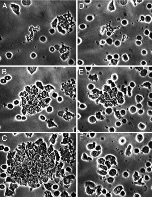
Fig. 2.
Reversal by oleic acid of the outsized chemotaxis induced by EDTA. Both preparations contain EDTA. (A–C) EDTA control cells, having received only the solvent for oleic acid (EtOH); (D–F) cells that received oleic acid. (A and D) Fields just after central erythrocytes have been destroyed by ruby laser microirradiation, creating a chemotactic gradient. (B and E) 15 min later: with EDTA alone (B), neutrophils continue to arrive; with EDTA + oleic acid (E), maximum aggregation has occurred and cells have begun to disperse (as normal cells do without EDTA). (C and F) After an additional 21.5 min, with EDTA alone (C), neutrophils are still arriving; with EDTA + oleic acid (F), neutrophil locomotion is normal but direction is random. EDTA, 5 mM; oleic acid, 125 μM; EtOH, 0.2%.

