Fig. 2.
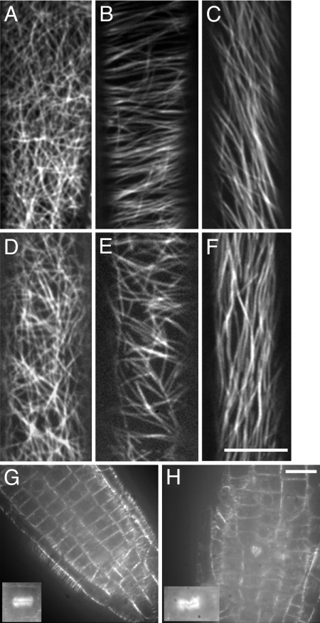
pfd6–1 affects microtubule organization in hypocotyl cells. GFP-MAP4-labeled microtubules in wild type (A–C) and pfd6–1 (D–F) hypocotyl cells. Images were collected at different positions of hypocotyl cells. Microtubule organization has distinct patterns for cells at the apical hook (A and D), 2–3 mm below apical hook (B and E), and 2–3 mm away from root (C and F). (Scale bar, 10 μm.) (G and H) Abnormal orientation of cell division planes in pfd6–1. Cells were visualized with GFP-MAP4 in roots of 3-day-old de-etiolated wild type (G) and pfd6–1 (H). An abnormal phragmoplast may lead to the cell plate defects observed in pfd6–1. (Scale bar, 10 μm.)

