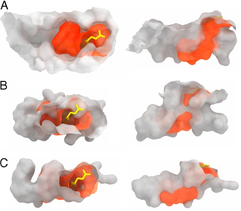
Fig. 4.
Molecular surfaces of MHC- and CD1-binding grooves. The chCD1–2 binding groove (orange) with bound palmitic acid is shown superimposed onto molecular surfaces of chicken MHC (A), hCD1d (B), and hCD1a (C). The left column represents a top view looking down onto the MHC- and CD1-binding grooves as in Fig 1B. The right column shows the binding pockets in a side view. Note that the chCD1–2-binding groove seems to be more similar to CD1a than to CD1d, but it is much smaller.

