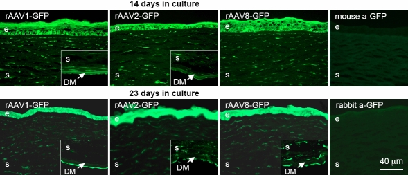
Figure 4.
rAAV-GFP transduction of organ-cultured human normal and diabetic corneas. Virus serotypes 1, 2, and 8 were used. Upper row – staining with mouse a-GFP MAB3580 (right panel – staining of an organ-cultured cornea not exposed to rAAV-GFP [negative control] with the same antibody [mouse a-GFP]); lower row – staining with rabbit a-GFP antibody ab6662 (right panel – staining of another negative control cornea with antibody ab6662 [rabbit a-GFP]). Epithelial (e), stromal (s), and endothelial (inset panels) cells are well transduced by rAAV-GFP. No staining is seen in corneas not exposed to the GFP-expressing virus. Both antibodies give very similar patterns. GFP was revealed by indirect immunofluorescent staining. DM, Descemet’s membrane. Bar=40 μm.

