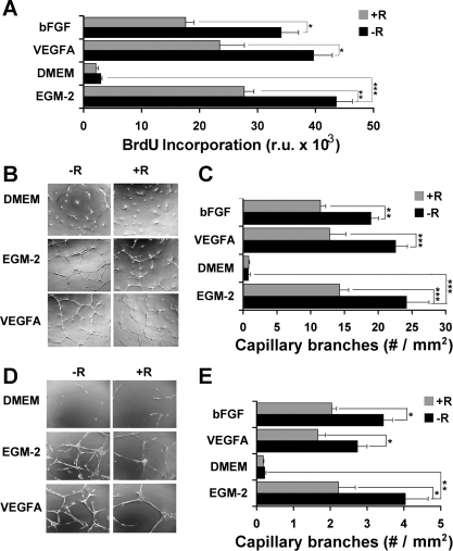
Fig. 4.
Analysis of the effect of RSG on endothelial cell proliferation and tube formation. A: cell proliferation in response to VEGFA or basic fibroblast growth factor (bFGF) in the absence (−R) or presence (+R) of 1 μM RSG was measured by BrdU incorporation, as described in materials and methods. Shown are means ± SE of 3 independent experiments performed in triplicate (total n = 9). Representative photomicrographs showing formation of capillary tubes by human umbilical endothelial cells (HUVEC; B) or microvascular endothelial cells (D) incubated for 24 h under the conditions indicated. Original magnification for HUVEC is ×40 and for microvascular endothelial cells is ×100. C and E: quantitative analysis of capillary tube formation by HUVEC (C) or microvascular endothelial cells (E). In each experiment, 4 fields of each of 3 wells/condition were analyzed, and the experiment was repeated 3 times (total, n = 36). Statistical analysis was performed by ANOVA followed by Tukey's multiple comparison test. *P < 0.05; **P < 0.01; ***P < 0.001. EGM-2, endothelial cell basal medium-2 supplemented with endothelial growth factor. RU, relative units.

