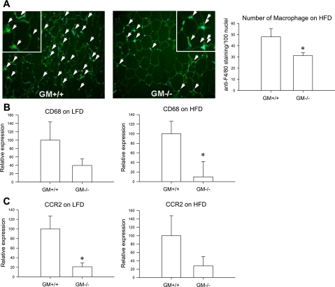
Fig. 1.
Representative figures of anti-F4/80 staining in mesenteric fat of wild-type mice and granulocyte-macrophage colony-stimulating factor (GM-CSF) knockout mice on a high-fat diet (HFD). White arrows indicate colocalization of F4/80 staining and 4′,6-diamidino-2-phenylindole (DAPI). No. of macrophages was determined by counting anti-F4/80 staining (bright green) per 100 nuclei (blue) in A. B: CD68 mRNA expression in mesenteric fat of wild-type mice (n = 6) and GM-CSF knockout mice (n = 4). C: CCR2 mRNA expression in mesenteric fat of wild-type mice and GM-CSF knockout mice on a low-fat diet (n = 7 and n = 8) or HFD (n = 6 and n = 5, respectively). *P < 0.05, wild-type mice vs. GM-CSF knockout mice.

