Fig. 8.
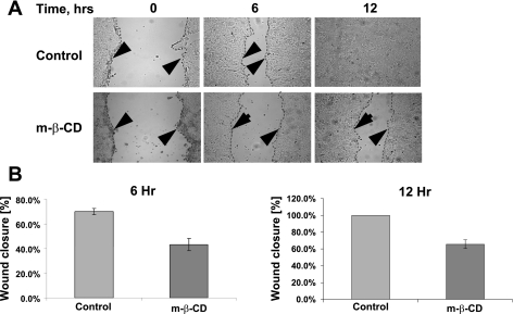
MβCD inhibits migration of IEC. A: confluent SK-CO15 cells were wounded and allowed to migrate for 6 and 12 h. MβCD (2.5 mM) or vehicle was added on the onset of cell migration. The dashed lines and the arrowheads highlight the leading edge of cells migrating in the wound. B: velocity of the wound closure is presented as a percentage of the initial wound widths. Note a significant attenuation of wound closure in the presence of MβCD (n = 4, P < 0.0004).

