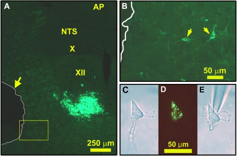
Fig. 1.
Retrograde labeling with fluorescent latex beads of XII premotor neurons and their identification after cell dissociation. A: fluorescein isothiocyanate (FITC)-labeled bead injection site in the ventral part of the XII nucleus and location of tissue micropunch taken from the medullary intermediate reticular (IRt) region (outlined in white and shown by arrow). B: expanded image of the region framed in yellow in A located just medial to the tissue punch; arrows point to 2 retrogradely labeled cells that remained in the slice. C–E: a cell dissociated from an IRt tissue punch and containing FITC-labeled beads, as seen under phase contrast (C), fluorescent illumination (D), and again phase contrast when the cell was attached to the tip of the sampling pipette (E). AP, area postrema; NTS, nucleus of the solitary tract; X, dorsal motor nucleus of the vagus; XII, hypoglossal motor nucleus.

