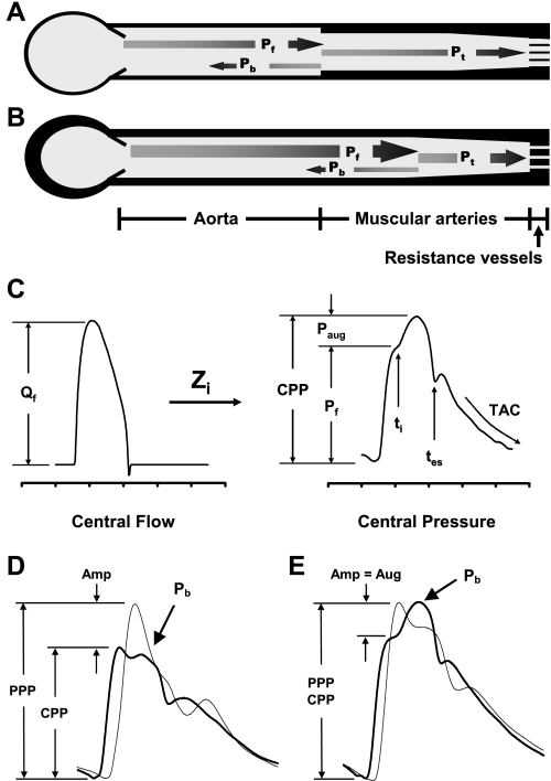
Fig. 1.
A brief synopsis of pulsatile hemodynamics. A: a simple model of the arterial system with a single dominant reflecting site at the interface between aorta and muscular arteries. When the forward wave (Pf) encounters the reflecting site, a portion is reflected (Pb) and most is transmitted (Pt). B: when the aorta stiffens (denoted by a thicker wall), impedance mismatch is reduced, the reflecting site shifts distally, a smaller fraction of Pf is reflected, and therefore more is transmitted into the microcirculation, which may trigger hypertrophic remodeling. C: the input flow wave interacts with input impedance (Zi) to produce the resulting pressure wave. The forward-flow wave (Qf) interacts with characteristic impedance (Zc) to produce Pf. Wave reflection and overlap between Pf and Pb creates late systolic augmentation (Paug), which contributes to central pulse pressure (CPP). The timings of wave reflection (ti) and systolic ejection (tes) are noted, and considerable overlap between Pf and Pb is evident (ti < tes). Total arterial compliance (TAC) can be computed by analyzing the pressure decay in diastole. PPP, peripheral pulse pressure. D: representative carotid (dark) and brachial (light) waveforms in a young healthy person. Note substantial amplification (Amp) of the pulse pressure with distal propagation. E: similar waveforms in an older individual with prominent central augmentation (Aug), which obscures normal peripheral amplification (Amp) of the forward-wave peak. [A and B reproduced with permission from Vyas et al. (96).]

