
Fig. 2.
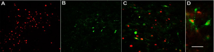
A subpopulation of FosB-positive neurons coexpresses corticotropin-releasing factor (CRF) in the PVN. A: immunofluorescence demonstrates RH-induced FosB expression (red). B: α-CRF antibody (green) identifies a subpopulation of neurons in the PVN and labels both cell bodies and fibers. C and D: successively higher (×40 and ×60) magnifications of merged images, respectively, of the same field double-labeled for both FosB and CRF exhibit colocalization of both antibodies in ∼30% of FosB-positive neurons. Scale bar represents 100 (A and B); 50 (C), and 25 (D) μm.