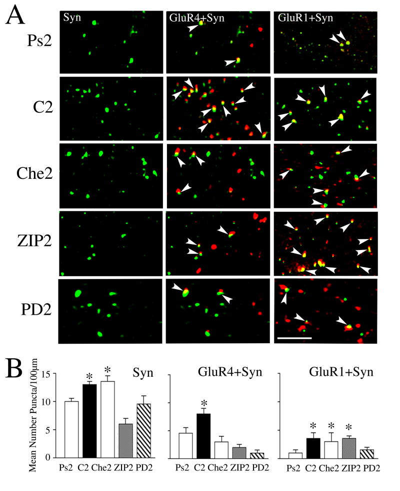
Fig. 8.
Colocalization studies show that synaptic incorporation of GluR4 AMPAR subunits is attenuated by inhibitors of PKC and ERK while GluR1 is attenuated by inhibitors of ERK. (A) Confocal images of selected abducens motor neurons from the different experimental groups showing punctate staining for synaptophysin (Syn), and colocalization of GluR4 subunits (GluR4 + Syn) and GluR1 subunits (GluR1 + Syn) with synaptophysin. Colocalization of AMPARs (red) with Syn (green) is defined by overlapping (yellow) or adjacent puncta and are indicated by the arrowheads. (B) Quantitative analysis of Syn, GluR4 + Syn, and GluR1 + Syn punctate staining for the different experimental groups is plotted. Preparations conditioned for two pairing sessions (C2) showed significantly greater punctate staining for Syn, GluR4 + Syn, and GluR1 + Syn compared to the pseudoconditioned group (Ps2). Treatment with the PKC inhibitor Che for two sessions (Che2) had no effect on the conditioning-induced increase in Syn or GluR1 + Syn, but significantly reduced GluR4 + Syn colocalization. Application of the PKCζ blocker ZIP for two sessions (ZIP2) significantly attenuated Syn and GluR4 + Syn, but not GluR1 + Syn. The MEK-ERK antagonist PD98059 (PD2) suppressed punctate staining for Syn, and colocalization of GluR4 + Syn and GluR1 + Syn. * indicate significant differences from Ps2. Scale bar = 2 μm.

