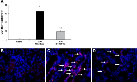
Fig. 4.
Macrophage infiltration. The number of CD11b+ cells in the outer stripe of the outer medulla in ischemic AKI was significantly reduced in IL-18BP Tg kidneys compared with WT kidneys (A). *P < 0.001 vs. sham. **P < 0.01 vs. WT AKI. Representative images of CD11b-positive staining in macrophages (arrows) in sham-operated mice (B), WT mice with AKI (C), and IL-18BP Tg mice with AKI (D) are shown.

