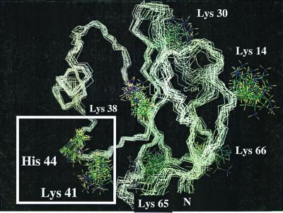
Figure 1.
NMR structure of the protein backbone of human CD59. The figure shows the 20 lowest energy structures of human CD59 with all lysine side chains and H44 (PDB Id: 1CDQ) (17). The structures were superimposed for the backbone of the β turn 41–44. The square highlights the K41–H44 glycation motif. K41 is within 5.91 ± 1.44 Å of the D1 imidazolic nitrogen of H44.

