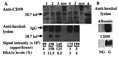
Figure 5.
Glycated CD59 in human urine. (A) Urine samples from diabetic (lanes 2 and 3) and nondiabetic (lanes 1, 4, and 5) subjects were concentrated by ultrafiltration and separated by anion exchange chromatography, and fractions were dot-blotted for the presence of CD59 with anti-CD59-specific Ab. CD59-positive fractions were pooled and immunoprecipitated with the HC1 anti-CD59-specific Ab. Immunoprecipitates were spun down and boiled for 30 min; then the immunocomplexes were separated by SDS/PAGE and immunoblotted with the monoclonal YTH53.1 anti-CD59 Ab (A, Upper), and with the anti-hexitol–lysine Ab (A, Lower). The Upper samples were separated by using a 20-cm-long gel that resolved the multiple CD59 bands (mw = molecular weight markers). The Lower samples were separated by using a minigel. Immunoaffinity-purified hRBC CD59 was included in the Upper gel as a control (lane 6). The signal intensity of the immunoblot bands marked by the arrows was quantified by BIOQUANT image analysis software. HbA1c levels were measured at the clinical laboratory of The Joslin Diabetes Center by HPLC. (B) Immunoblot of glycated (G) and nonglycated (NG) albumin (Upper) and immunoaffinity-purified hRBC CD59 (Lower) with anti-hexitol–lysine Ab.

