Fig. 2.
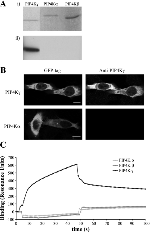
Specificity of anti-PIP4Kγ antibody. Detection of PIP4Kγ, but not the PIP4Kα or PIP4Kβ isoforms, was established in various procedures. A: equivalent amounts of purified recombinant protein of each isoform were visualized on SDS-PAGE (i) and detected by Western blotting (ii). B: HeLa cells overexpressing green fluorescent protein (GFP)-tagged PIP4Kγ or PIP4Kα were fixed and stained with anti-PIP4Kγ antibody. Scale bar = 10 μm. C: Surface plasmon resonance analysis using PIP4Kα, PIP4Kβ, and PIP4Kγ adsorbed to an nitrilotriacetic acid biosensor. Anti-PIP4Kγ antibody was passed over the biosensor at 0 s and exchanged for wash buffer at 45 s. Data were normalized to 6xHis tag control protein binding.

