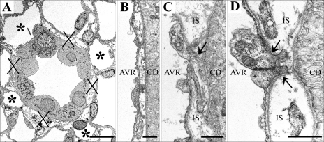
Fig. 11.
Electron micrographs showing transverse sections of CDs and AVR from ∼1.5 (A, B, and D) and 4 mm (C) below the base of the IM. A: CD surrounded by 4 AVR (asterisks). Other tubular structures surrounding the CD are ATLs. Interstitial nodal spaces (X) are formed between CD, AVR, and ATLs. Scale bar, 10 μm. B: AVR abuts CD with minimal direct contact. Scale bar, 1 μm. C: AVR abuts CD with microvillus (arrow). IS, interstitium. Scale bar: 1 μm. D: AVR abuts CD with microvilli (arrows). Scale bar, 1 μm. From Pannabecker and Dantzler (53).

