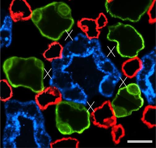
Fig. 12.
One single CD in transverse section showing interstitial nodal spaces (X) between CD (blue), AVR (red), and ATLs (green) in a composite image of 2 sections 3 μm apart, from near the IM base. Open space in wall of central CD is the location of an intercalated cell, which does not label for AQP2. Scale bar, 10 μm. From Pannabecker and Dantzler (53).

