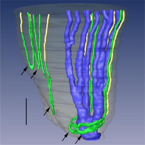
Fig. 13.
Three-dimensional reconstruction of several papillary CDs (AQP2, blue), ATLs (ClCK-1, green), and AQP1-null DTLs (AQP1 and α-B crystallin, yellow). Papillary surface epithelium is shown in gray. Tight narrow bends of loops of Henle (top 3 arrows) and wide transverse bends of loops of Henle (bottom 2 arrows) are shown. Wide transverse bends of 2 loops reaching to near the tip of the papilla almost completely encompass a final CD segment (blue) before its merging with the papillary wall (surface epithelium; translucent gray) to form a duct of Bellini. Relative diameters of loops and CDs in this image nearly approximate true dimensions. Scale bar, 200 μm. From Pannabecker and Dantzler (54).

