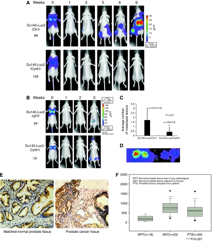
Figure 6.
Silencing of Cyr61 eliminated the systemic tumour growth of Du145 cells in vivo. (A, B, C, and D) Either Du145-Luc2/Ctrli #1 or Du145-Luc2/Cyr61i #1 cells (1 × 10⁷) and 5 × 10⁶ of either Du145-Luc2/GFP or Du145-Luc2/Cyr61 cells were injected into each 10 mice tail vein separately. The bioluminescence images were acquired using the IVIS imaging box at indicated time points. Representative mice (8# and 10#) of Du145-Luc2/Ctrli #1, Du145-Luc2/Cyr61i #1 (A) and Du145-Luc2/GFP, Du145-Luc2/Cyr61 (B) were presented for each time point. (C) Average number of metastasis lesions was qualified. (D) The metastasis lesion was picked out to confirm that bioluminescence signalling is not artificial. (E) Immunohistochemistry of Cyr61 in clinical prostate cancer samples and matched normal prostate tissue. Scale bar=100 μm. (F) Elevation of Cyr61 level upon tissue adjacent to tumour and the tumour samples compared with normal prostate tissues free of any pathological alteration; box-and-whisker plots of Cyr61 expression level at different stages of prostate cancer tissues. ***P<0.001; **P<0.01.

