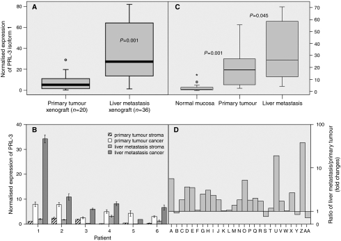
Figure 1.
(A) Normalised expression of PRL-3 in xenografts determined by quantitative –PCR. Boxplots comparing PRL-3 mRNA expression in 20 human colorectal primary tumours and 36 liver metastases from CRC raised as xenografts in nu/nu Swiss mice. (B) Normalised expression of PRL-3 determined by Quantitative –PCR in paired samples of microdissected specimens. We analysed four different cell populations from six different patients: (i) primary tumour stromal cells, (ii) primary tumour cancer cells, (iii) hepatic metastases stromal cells and (iv) hepatic metastases cancer cells, obtained by means of Arcturus PixCell II Laser-Capture microdissection system. (C) Boxplots comparing PRL-3 mRNA normalised expression in 27 matched fresh-frozen samples of non-adjacent normal mucosa, primary tumour and liver metastases (22 synchronous and five metachronous). Comparing normal colonic mucosa and primary tumours, pair-wise fixed reallocation randomisation test (Pfaffl, 2001) showed significantly higher expression in carcinomas by the factor 23.133 (P<0.001). Besides, liver metastases displayed a higher expression than primary colorectal tumours (factor 1.983; P=0.045). (D) Bars displayed the overexpression of isoform 1 PRL-3 in 23 out of 27 liver metastases. Bars represent the ratio among PRL-3 normalised expression in liver metastases divided by PRL-3 normalised expression in its paired primary tumour. The ratio was expressed as fold changes. In all experiments, the level of PRL-3 expression was expressed as absolute expression normalising for β2-microglobuline as housekeeping control gene. All experiments were performed in triplicate using two different retrotranscriptions.

