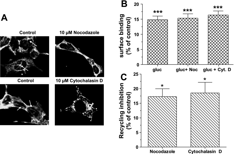
Fig. 4.
Role of cytoskeleton in GR internalization and recycling. A: HEK-GR cells were treated with either 10 μM nocodazole or 10 μM cytochalasin D for 30 min at 37°C. The cells were permeabilized and immunostained with either the mouse anti-β-tubulin antibody or the mouse anti-β-actin antibody. The arrows indicate starlike structures of microtubules (top) and actin filaments (bottom). The images shown are representative of 3 independent experiments: B: HEK-GR cells were treated with either 10 μM nocodazole (Noc) or 10 μM cytochalasin D (Cyt D) for 30 min at 37°C, followed by 30-min incubation in the absence (control) and presence of 100 nM glucagon at 37°C. Surface binding was determined by radioligand binding assay (method B). The data shown are means ± SE of 3 independent experiments. C: after glucagon treatment, the cells were washed and incubated without (control) and with either 10 μM nocodazole or 10 μM cytochalasin D and returned to 37°C for 30 min. Surface binding was determined by radioligand binding assay (method B). Data represent means ± SE of 3 independent experiments. Data were analyzed by one-way ANOVA and Bonferroni posttest. *Significantly different from control: P < 0.05. ***Significantly different from control: P < 0.001.

