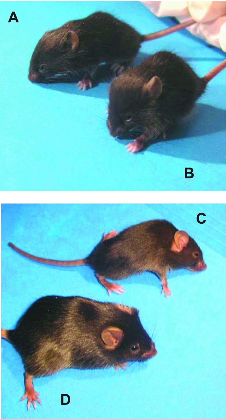
Fig. 1.
Two- and 6-wk-old control mice (A and D) and age-matched mice with muscular dystrophy with myositis (mdm; B and C). At 2 wk, mdm mouse was phenotypically indistinguishable from its wild-type littermate (A and B). Progression of disease was marked at 6 wk, manifesting in severe wasting of hindlimb muscles, dorsal kyphosis, decreased ambulation, and decreased body mass (C).

