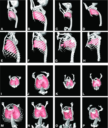
Fig. 2.
Three-dimensional surface reconstructions of thoracic skeleton and lungs from high-resolution micro-CT. Images are representative of control and mdm mice at 2 and 6 wk of age (n = 3 for each genotype and age group). A, E, I, and M (column 1) represent wild-type mice for comparison with images of mdm mice (columns 2, 3 and 4). Sagittal views are shown for 2-wk-old (A–D) and 6-wk-old (E–H) mice. Corresponding axial views are also shown for the same 2-wk-old (I–L) and 6-wk-old (M–P) mice. Note normal spinal curvature, symmetric thoracic rib cage configuration, and normal lung shape in 2-wk-old mdm compared with wild-type mice. Note severe scoliosis and dorsal kyphosis, in combination with asymmetric distortion of the thoracic rib cage, namely, lateral constriction and anterior-posterior lengthening, in addition to decreased skeletal and lung mass in 6-wk-old mdm mice. Lung shapes in 6-wk-old mdm mice and appeared to be distorted concomitantly with rib cage changes and appeared smaller than controls.

