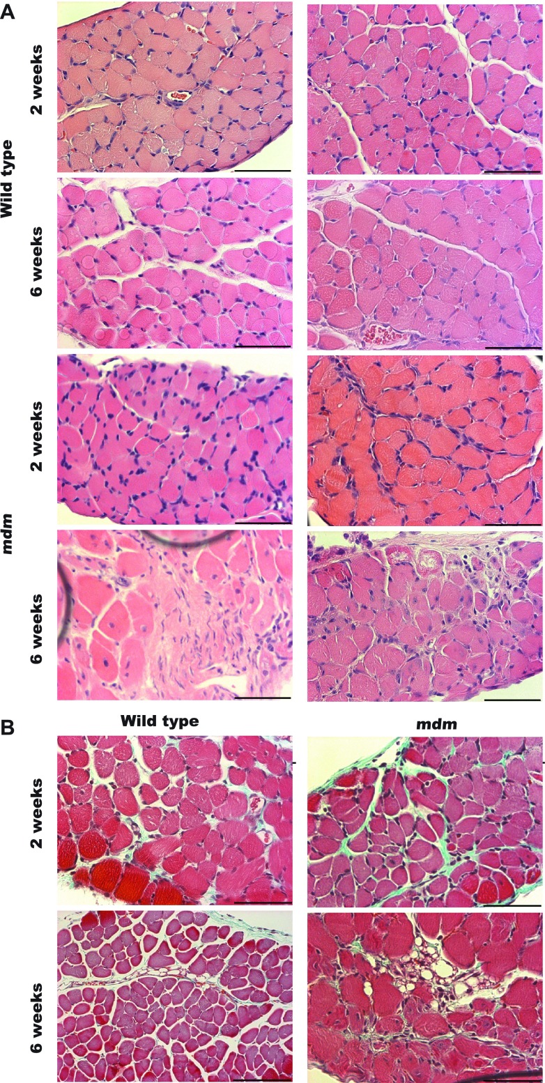
Fig. 5.
Histological staining of 2- and 6-wk diaphragms. A: hematoxylin-eosin staining of wild-type and mdm diaphragms (n = 3 for each age × genotype group) at 2 and 6 wk. Rows 1 and 2 are images of wild type diaphragms. Rows 3 and 4 are images of mdm diaphragms. Columns 1 and 2 show examples of the same age × genotype groups. At 2 wk, histopathological signs were largely absent in mdm diaphragm. At 6 wk, mdm diaphragms showed marked foci of necrosis, scar tissue, and variation in myofiber size. B: small foci of increased perimysial connective tissue deposition in 2-wk mdm diaphragm stained with Masson's trichrome. At 6-wk, large endomysial and perimysial connective tissue deposition was noted in localized foci of diaphragm necrosis. Scale bar, 60 μm.

